Estimation of Endocranial Capacity and Identification of Sex from Adult Human Skull of Eastern India
Dr. Kanhu Charan Satapathy
Bidya Bharati Sahoo
| Abstract | Observations and Results |
| Introduction | Discussion |
| Materials and Methods | Conclusion |
Abstract
Endocranial capacity is an important measurement in the study of human evolution, variation and for clinical practice for the study of abnormalities of cranial size and shape. The cranial capacity gives a reasonable estimate about the volume of brain. In the present study an attempt has been made to measure endocranial capacity of the 83 dry human skulls preserved in the department of Anthropology, Utkal University, Bhubaneswar, Odisha, during the year 2013-2014. Skulls choosen for the present study were of adult age only. Dry mustard seeds of uniform size were use to fill cranial cavity to determine the capacity. Mean cranial capacity of male skull was found to be 1329.42, while in female skull mean cranial capacity found to be 1235.61. A highly significant difference (p<0.01) was observed between intracranial volumes of male and female skulls when compared. The present findings are expected to be of forensic anthropological and clinical importance for eastern Indian population.
Key words : Endocranial capacity, sex determination, Human skull
1. Introduction:
Determination of sex is an important criterion, for identification of an individual for medico-legal purposes. Skull and pelvis assumes great importance in establishing sex of an individual (Lalwani et.al, 2012). Various dimensions and indices have been reported as valuable indicators in the differentiation of male and female skulls. Knowledge of the volume of cranial cavity of either a dry skull or of a living being may be important for the study and comparison of the crania of populations with various fundamental differences like racial, geographical, ethnic, dietary, environmental etc (Manjunath, 2002a). Endocranial capacity is widely used as a substitute for actual brain size, which is one of the most important variables for human evolution (Muralidhar et.al, 2014). It is well accepted that the cranial capacity, which is in close correlation with brain volume, reflects the racial characteristics, and this has been considered as one of commonest topics in physical anthropological studies (Hwang et al; 1995, Manjunath, 2002a, Wolf et al., 2003; Mazonakis et al., 2004). The skeletal maturity in case of males and females grows at different rates during their growth (Krogman, 1978). The female skull has a capacity about one tenth less than that belonging to male of the same race (ibid).The cranial capacity of female is about 10-11% less than male is mostly reflecting large male body mass(Bannister et al,1995). Hence it can be used as a parameter for sex determination. It can be correlated with other cranial parameters and in the study of primate phylogeny. The endocranial capacity of modern man ranges between 1200-1800cc but largely it ranges between 1350-1400cc (Hwang et al., 1995). The endocranial capacity in the first few months of life attains on average 900cc in males and 600cc in females. By 2 years of age endocranial capacity rises to 1150cc in males and 1000 cc in females, whereas by 5 years it rises to 1350cc in males, 1200cc in females, which, respectively are 77% and 90% of the endocranial capacity observed at age of 15 years, this capacity at this age being 1500cc in males and 1300cc in females (Martin, 1981). The present paper reports the determination of sex from cranial capacity of dry human skulls preserved in the museum of the Department of Anthropology, Utkal Univesrity, Bhubaneswar, Odisha.
2. Materials and Methods:
The craniometric study was undertaken during the year 2013-2014. All the skulls used for the study were those of adults and the number of such skulls was 83. The endocranial capacity of dry human adult skulls was measured by manual packing method (Mac Donell, 1904; Hardilica, 1920; Steward, 1934 ; Manjunath, 2002). The packing material used in the present study was mustard seeds instead of usual millet seeds obtained in the local market. Only the intact undamaged skulls with unknown sex and without any injury, pathology or congenital anomaly were selected, for the study. While taking the measurements each one was taken to the nearest millimetre at least three times and the average was considered for calculation (Singh and Bhasin, 1968). The data of each crania were recorded and analysed by IBM SPSS 20.0 and MS Excel 2007. From morphological characters (Appendix-1, Jain et al, 2013) of these 83 dry skulls it was found that 55 of these skulls were of males while rest 28 were of females. In estimating the cranial volume of these 83 skulls the following methods were followed.
2.1 Manual Packing Method (Steward, 1934):
The following steps are followed in this method.
Step-1 The optic canals, supraorbital and infraorbital fissures were plugged with cotton.
Step-2 The foramina in the base of the skull except the foramen magnum and small foramina less than the size of mustard seed were plugged with cotton.
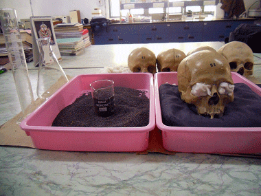
Step-3 The skulls were placed on a cotton cushion kept at the center of a tray, with foramen magnum upwards and its panel parallel to the ground.
Step-4 Then mustard seeds were poured into skull cavity through foramen magnum and skull was shaken vigorously with hands, so that the seeds settle into frontal part of the skull. The cavity is filled up with more seeds up to the rim of the foramen magnum while shaking the skull from time to time. Wooden stick was pushed through the foramen magnum into the mass of the seeds, in different directions. Finally the seeds were pressed gently with thumb at the foramen magnum. This process was repeated until the entire cavity was filled up with seeds and no more seeds were required to be poured into it.
Step-5 After the skull was fully filled the seeds were poured from the skull into a glass jar and measuring cylinder. The volume of these seeds was measured from the measuring cylinder, which in turn determined the cranial volume.
2.2. Linear Measurements :
By this method the measures of the three principal dimensions of the cranium, namely, (1) Maximum anterior-posterior length (L), (measured between Glabella and the Inion).(2) Maximum breadth(B), ( biparietal diameter; measured between two parietal eminences Euryon ).and (3) Cranial height(H) (basi- bregmatic height, measured between the internal acoustic meatus to the highest point of the vertex i.e. the Bregma ) were obtained. And from these dimensions the cranial volume was calculated. The formulas used were as follows
2.2.1. Lee and Pearson (1901) Formula : According to this formula the cranial volume is for
Males: 359.34+0.000365*L*B*H
and
Females: 296.40+0.000375*L*B*H
2.2.2. Dekaban and Lieberman (1964) Formula : According to this formula the cranial volume is 0.5238*L*B*H.:
2.2.3 Williams et.al. (1995) and Manjunath (2002b) Formula : According to this formula the cranial volume is for
Males: 0.000337 (L-11) (B-11) (H-11) +406.01 cc
and
Females: 0.000400 (L-11) (B-11) (H-11) +206.60 cc
2.3 Classification of skulls : Size of the skull may be classified according to their cranial capacities. Henry Gray ( 2000), and Brash (1951) reported that the size of the skull varies considerably in different races of man. The skulls of carnial volume less than 1350cc are called Microcephalic skulls, the skulls whose carnial volume lies between 1350cc and 1450 cc, both the end values inclusive, are called Mesocephalic skulls, where as the skulls of carnial volume greater than 1450cc are called Megacephalic skulls
3. Observations and Results:
3.1 Cranial Capacity of the Skulls .
In the present study, cranial capacity, determined by manual packing method, for 55 male skulls, identified morphologically (Appendix-1), lied between 1312.38-1444.25cc so that the mean turns out to be 1329.42cc. For rest 28 female skulls, it was found to be between 1236.15-1084.00cc with a mean of 1235.61cc. Thus, a significant difference between male and female skulls was observed. (Table 1).
Table 1: Range, mean, standard deviation of endocranial capacity .
|
Sex of dry human skull |
N |
Range |
Mean |
Standard deviation |
|
Male |
55 |
1312.38-1444.25c.c |
1329.42c.c |
154.38 |
|
Female |
28 |
1236.15-1084.00c.c |
1235.61c.c |
135.94 |
F (1, 83) = 7.406, p <0.01
On analysis of the cranial volumes of the 83 skulls under study, the F-value turns out to be 7.406 and the corresponding p-value is 0.008 at one degree of freedom. The difference observed between male and female for cranial capacity is found statically significant.
But the linear measurement methods led to different mean values of cranial capacity for the skulls under study ((Table-2)).. It is only the formulas of Manjunath (2002b) and Williams et.al (1995), which gave values of cranial capacity close to the values obtained by manual packing method of Stewards (1934). Other formulas, namely, the formula of Dekaban and Liberman(1964) and the formula of Lee and Pearson (1901) gave much higher values.
Table 2: Cranial capacity using formulas of different scholars .
|
Sex of dry skull |
Dekaban & Liberman, 1964 |
Lee –Pearson, 1901 |
Manjunath, 2002b & Williiams et.al.1995 |
Manual packing method Stewards, 1934 |
|
Male |
1762.97c.c |
1558.55c.c |
1367.80c.c |
1329.42c.c |
|
Female |
1607.21c.c |
1447.03c.c |
1250.54c.c |
1235.61c.c |
The determination of cranial capacity of skulls has been attempted by many authors at different points of time, from different regions of the world with different sample sizes, which is presented below (Table 3)
Table 3: Values of cranial capacity of human skulls by different scholars of different regions of world.
|
Sl No. |
Name of workers |
Place & country of work |
Year of work |
Sample size of study |
Cranial capacity of male in cc |
Cranial capacity of female in cc. |
|
1 |
Ricklan et al |
Zulu skull (South Africa) |
1986 |
50 males & 50 females |
1373.3 |
1251.0 |
|
2 |
Hwang et al |
Korean |
1995 |
64 males & 23 females |
1470.0 |
1317.0 |
|
3 |
Manjunath KY |
Bangalore, India |
2002 |
33 males & 17 females |
1152.81 |
1117.82 |
|
4 |
Golalipour MJ et al |
North Iran (Native Fars ) |
2005 |
198 males & 203 females |
1369.60 |
1215.40 |
|
5 |
Golalipour MJ et al |
North Iran (Turkman) |
2005 |
200 males & 207 females |
1420.60 |
1227.40 |
|
6 |
Gohiya VK et al |
Madhya Pradesh, India |
2010 |
200 males &200females |
1380.52 |
1188.75 |
|
7 |
Mania MB et al |
Maiduguri, Nigeria |
2011 |
150 males &150 females |
1424.4 |
1331.3 |
|
8 |
Salve VM et al |
Andhra region, India |
2012 |
160 males & 160 females |
1322.78 |
1112.49 |
|
9 |
Lalwani M et al |
Bhopal(M.P),India |
2012 |
100males & 60 females |
1302.95 |
1179.92 |
|
10 |
Present study |
Odisha, India |
2014 |
55 males & 28 females |
1329.42 |
1235.61 |
3.2. Classification of Skulls
The skulls under study were classified according to the prescriptions of Henry Gray (2000) & Brash (1951), which is presented as follows (Table 4) .
Table 4: classification of skulls based on cranial capacity (Henry Gray, 2000 & Brash, 1951).
|
Sex |
Microcephalic (< 1350 cc) |
Mesocephalic (1350-1450cc) |
Megacephalic (>1450cc) |
Total |
|
Male |
33(60.0) |
12 (21.82) |
10(18.18) |
55(66.27) |
|
Female |
22(78.57) |
5(17.85) |
1(3.57) |
28(33.73) |
|
Total |
55(66.27) |
17(20.48) |
11(13.25) |
83(100.00) |
3.3. Correlation coefficients between cranial capacity and cranial dimensions
For the skulls under study, the Correlation coefficients between cranial capacity and all measured cranial dimensions were found to be statistically significant and positive indicating a strong relation between the above parameters.(Table 5)
Table 5: Correlation coefficient between the cranial capacity & cranial dimensions of male and female.
|
Parameters |
Present study on dry Crania2014 |
Ilayperuma.2011(20-23yrs adult Sri Lankan Population) |
||||
|
Male |
female |
both sexes |
Male |
female |
both sexes |
|
|
Cranial capacity & cranial length |
0.753 |
0.462 |
0.709 |
0.806 |
0.255 |
0.654 |
|
Cranial capacity & cranial breadth |
0.606 |
0.489 |
0.601 |
0.663 |
0.839 |
0.780 |
|
Cranial capacity & auricular head height |
0.674 |
0.303 |
0.607 |
0.882 |
0.884 |
0.835 |
3.4. Sexual Dimorphism Index of cranial capacity:
The sexual dimorphism index of cranial capacity is determined by the formula
Male mean capacity- Female mean capacity
= ------------------------------------------------------- X 100.
Male mean capacity
For the present study, when male mean cranial capacity is 1329.42cc and female mean cranial capacity is 1235.61 cc, sexual dimorphism index turns out to be 7.05.
4. Discussion:
In Forensic investigation, sex determination is a necessary requirement for identification of skeletal remains in their advanced state of decomposition. The present study has clearly shown (Table-1) that the cranial capacity of male skulls is significantly different from that of female skulls. The mean cranial capacity of male skulls (55) is 1329.42±154.38 c.c ( range 1312.38-1444.25c.c), whereas the mean cranial capacity of female skulls(28) is 1235.61±135.94 c.c (range 1084.00-1236.15c.c).
According to some Indian studies the cranial volume of adult crania has been found to very between 950c.c-1520c.c (Shukla AP, 1966, Routal RV, 1984). Pal, Bhagawat and Routal (1986) in their further studies of 370 Gujurati(Indian) crania have estimated the cranial volume as ranging from 1030-1620 c.c(Mean: 1252 ±112.84)). According to Hwang et.al (1995) report, the cranial volume was 1470±117 c.c in female skull. Golalipour et.al (2005) reported cranial capacity of the Turkman was 1420.60±85 c.c in male and 1227.2±120 c.c in females, and in native fars group in male and female were 1369.4±142 c.c and 1215.8±125c.c respectively
As regards the classification of skulls (Table-4), it is interesting to find that almost two thirds of the skulls under study belonged to Microcephalic class and about one fifth belonged to Mesocephalic class and the rest belonged to Megacephalic class. Further comparison between male and female skulls belonging to these classes, it may be observed that the percentage of female skulls is quite higher than the percentage of male skulls in Microcephalic class, which is otherwise in other two classes. This suggests that Microcephalic class, that is the class of skulls of cranial capacity less than 1350cc, contains more human skulls than other classes. And between male and female skulls, a larger proportion of female skulls belong to this class than the proportion of male skulls. Study in respect of cephalic index in Odisha shows that there is not much divergence among the groups, and majority of them lie in the border line of dolichocephalic (long headed) and mesocephalic (medium headed) class (Basu et al 1995).
Sexual dimorphism is a vital component of the morphological variation among biological population (Gray Henry, 2000, Ilayperuma, 2011). The index of sexual dimorphism in the present study was 7.05%, which is less than that of the Sri Lankans (8.46%), Caucasians (7.95%), the Zulu(8.9%), the Koreans (10.3%), Tukmans (13.61%), Native Fars (11.22%) and Turkey population (10.06%) (Ricklan et al, 1986, Hwang et al 1995; Golalipour et al., 2005; Acer et al., 2007). According to Frayer and Wolpoff (1985) the sexual dimorphism in size, shape and behaviour does not occur in infants, children and sub adults, but are typical primarily in the adult stage, which, indicates that many of the effects are the result of hormonal events occurring at puberty. Similarly, according to Larsen (2003) the modern day homo sapiens exhibit relatively narrow range of sexual dimorphism with average body mass difference between sexes being roughly equal to 15% (compared to most primates and anthropoids, ranging 50-55%). The sexual differences in cranial dimensions of adults emphasize the significance of applying the anatomical variation data to an individual subject in a given population.
5. Conclusion:
The result of present study on human dry crania in eastern part of India shows the mean cranial capacity of human dry skulls is 1297.77c.c, which is grouped under microcephalic. The mean endocranial capacity of male skulls is higher than female skulls. Using proper statistical methods the difference in the cranial capacity of male and female skulls was found to be highly significant. Thus it can be inferred that the cranial capacity is a reliable craniometric method for sex identification.
References:
-
Acer, N., Usanmaz, M. Togay, U. Ertekin, T. Estimation of cranial capacity in 17-26 years old university students. International journal morphology ,2007; volume 25( 1),pages 65-70.
-
Bannister LH, Berry MM, Collins P, Dyson M et al. Grey;s anatomyical basis of medicine and surgery. 38th ed. Edinburgh: Churchill Livingstone, 1995: 576-568.
-
Basu, A., Sreenath I. Anthropometric variation in Assam, Bihar, & Orissa, Anthropological survey of India, 1995.
-
Brash, JC. Cunnigham’s Taxtbook of Anatomy, 9th ed. New York University Press, 1951.
-
Dekaban, A. and Lieberman , J.E., Calculation of cranial capacity by linear dimensions,
-
Anatomical Record , 1964; 115: 215-219.
-
Frayer DW and Wolpoff MH. Sexual Dimorphism. Ann.Rev. Anthropol. 1985, 14:429-473.
-
Gohiya VK, Shrivatava. S. & Gohiya, S., Estimation of cranial capacity in 20-25 year old population of Madhya Pradesh, a State of India. International journal of Morphology, 2010, Volume 28, Issues 4,pages 1211-1214.
-
Golalipour, M. J.: Jahanshaei. M. & Haidari , K . Estimation of cranial capacity in 17-20 years old in South East of Caspian sea border. Int. J. Morphology., 2005, 23(4) : 301-304.
-
Grey H. Grey’s Anatomy: The anatomical basis of medicine and surgery: 37th Edition. New York,
-
Churchill Livingstone, 2000, Pp 337-98.
-
Hawang Y, Lee KH, Choi BY, Lee KS, Lee HY, Sir WS, Kim HJ, Koh KS, Han SH, Chang MS and Kim H. Study on the Korean Adult Cranial Capacity. Journal of Korean Medical Science, 1995 August: 10(4):239-42.
-
Hardlica A. Anthropometry. Wister Institute, 1920,Philadelphia, .
-
Ilayperuma I, Cranial capacity in an adult Sri Lankan population : Sexual dimorphism and Ethnic Diversity, International J. Morphology, 2011, volume 29(2), page 479-484.
-
.Jain S.K, Choudhary A.K, Mishra P. Morphometric Evaluation of Foramen magnum for sex determination in a documented North Indian sample. Journal of Evalution of Medical and Sciences 2013,Volume 2, Issue 42, pages 8093-8098.
-
Krogman WM. The human skeleton in Forensic Medicine, 3rd Edition, Springfield, Thomas 1978, Pp 277-87.
-
Lalwani M , Yadav J, Arora A, Dubey BP. Sex identification from Cranial Capacity of Adult Human
-
Skulls, J Indian Acad Forensic Med., April- June 2012, volume-34, No. 2.
-
Larsen CS. Equality for the sexs in human evolution; early hominids sexual dimorphism and implications for mating systems and social behaviour. PANS, 2003; 100(16):9103-9104.
-
Lee A and Pearson K. Data for the problem of evaluation of the human skull – a first study of the correlation of human skull. Philosophical Transactions of Royal Society. London,1901, 196a:225-264.
-
MacDonell WR. A study on the variation and correlation of the human skull with special reference to English crania. Biometric, 1904, 3: 191-244.
-
Maina MB, Shaup YC, Garba SH, Muhammad MA, Garba AM, Yao AU, Omoniyi ON, Assessments of Cranial Capacities in a North –Eastern Adult Nigerian Population. Journal of Applied Science 2011; 11:2662-2665.
-
Martin, R. Relative brain size and basal metabolic rate in terrestrial vertebrate. Nature, 1981,293:57- 60
-
Mazonakis TM, Karampekios S, Dailakis J, Viloudaki A & Gourtsoyiannis N. Stereological estimation of total intracranial volume on CT images, Eur. Radial, 2004; 14:1285-90.
-
Murildhar P. S, Magi M, Nanjundappa B, Pavan P Havaldar, Premalatha Gogi, Sahik Hussain Saheb.
-
Morphometric analysis of endocranial capacity. Int J Anat Res , 2014, Volume2, Issue 3, pages 242-48,.
-
Manjunath, KY. Estimation of Cranial Volume an Overview of methodologies, Journal of Anatomy society India ,2002a ,volume 51,Issue 1, pages 85-91.
-
Manjunath, KY. Estimation of cranial volume in dissecting room cadaver. J Anat Soc Ind. 2002b; 51:168-72.
-
Pal GP, Bhagwat SS,& Routal RV. A study of sutural bone in Gujurati (Indian) crania. Anthropoligischer Anzeigher,1986;volume 44,Issue 1,pages 67-76.
-
Routal. R. V: Pal , G.P & Bhagwat, S.S relationship between endocranial volume and the area of the foramen magnum. J. Anat. Soc. India., 1984,volume 33,pages 145-149.
-
Recklan DE, Tobias PV. Unusually low sexual diamorphism of endocranial capacity in Zulu Cranial series. Am J Phys Anthrop 1986: 71(3):283-93.
-
Salve V.M, Gitte R.N, Estimation of cranial capacity of coastal Andhra Pradesh of India, Dr pinnamaneni sirdhartha Institute of Medical sciences & Reserch foundation, Chinnaoutpalli.Volume 10, Issue 3,pages 175-180,September-December 2012.
-
Shukla AP. A study of cranial capacity and cranial index of Indian skull. J. Anat. Soc. India , 1966, voume15,pages: 31-35
-
SinghIP & Bhasin MK , A laboratory manual on Biological Anthropology. Kamala-Raj- Enterpries, Delhi, 1968
-
Steward TD. Cranial capacities studies. Am. J Phys Anthro. 1934; XVIII(3):337-361.
-
Wolf H, Kruggel F, Hensel A, Wahland LO, arendt T & Gertz HJ. The relationship between head size and intracranial volume in elderly subject. Brain Res, 2003; 973:74-80.
-
Williams PL, Bannister LH, Dyson M, Collin Dussek JE and Ferguson JWM. Gray’s Anatomy, 39th ed. Edinburgh, London: Churchill Livingstone; 1995:487-489.
Appendix-I
Estimation of sex from the skull through morphological characters .
Morphological variation between male & female skull (S.K. Jain, 2013)
|
SL.N |
Traits |
Male |
Female |
|
1 |
General size |
Large |
Small |
|
2 |
Architecture |
Rugged |
Smooth |
|
3 |
Supraorbital ridge |
Medium to large |
Small to medium |
|
4 |
Mastoid process |
Medium to large |
Small to medium |
|
5 |
Occipital area |
Muscle lines and protuberance marked |
Muscle lines and protuberance marked not marked. |
|
6 |
Frontal eminence |
Small |
Large |
|
7 |
Parietal eminence |
Small |
Large |
|
8 |
Orbits |
Squared, lower, relatively smaller |
With rounded, higher, relatively larger, with sharp margins |
|
9 |
Forehead |
Steeper, less rounded |
Rounded, full, infantile |
|
10 |
Cheek bones |
Heavier, more laterally arched |
Lighter, more compressed |
|
11 |
Mandible |
Larger, higher symphysis, broader ascending ramus |
Small with less corpal and ramus dimensions |
|
12 |
palate |
Larger, broader, tends to U-shape |
Small and tends to parabola |
|
13 |
Occipital condyles |
Large |
Small |
|
14 |
Teeth |
Large, lower M1 more often 5 cusped |
Small, more often 4 cusps. |

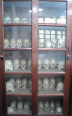
Plate 1: Dry human crania’s, Biological Anthropology Laboratory, Department of Anthropology, Utkal University,Bhubaneswar, Odisha

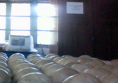
Plate2: Dry human crania considered for the present study.

Plate 3: Measurement of Cranial Volume

Plate 4: Measurement of Cranial Volume manually

Plate 5: Sliding Calliper and Spreading Callipers used in the present study.
----------------------------------------------------
Reader, P. G. Dept. of Anthropology
Research Scholar, Dept. of Anthropology, Utkal University, Bhubaneswar-4, Odisha


