Significance of Morphometric analysis of Foramen Magnum of Human Dry Crania for the Estimation of Sex
| Dr. Kanhu Charan Satapathy | |
| Bidya Bharati Sahoo |
| Abstract | Result and Discussion |
| Introduction | Conclusion |
| Materials and Methods |
Abstract
Forensic study is very important in the identification of sex especially when the body of the deceased, death being caused by physical injury due to weapons, fire or strong chemicals, is found in a decomposed state. The morphological characteristics obtained by craniometry may be the key to sex determination, which enable us to identify unknown individuals anywhere in the world. In the present study an attempt has been made to evaluate the effectiveness of the linear morphometry of foramen magnum to verify the morphological characteristics in dry human skulls for gender determination. Eighty-three dry human skulls of adult age used in the study belong to the museum of the Department of Anthropology, Utkal University, Bhubaneswar, Odisha. Length and Breadth of foramen magnum are measured in standard technique by vernier calliper. It is found that for male the mean value of LFM, BFM, Foramen magnum index, the area of foramen magnum (Teixeira) and the area of foramen magnum (Routal) are respectively 3.65c.m, 3.12c.m, 85.48, 9.00 and 8.94, whereas the corresponding values for the female are 3.52c.m, 2.98cm, 84.66, 8.29 and 8.23. The difference in length, breadth and area of foramen magnum between male and female was found to be statistically significant (p<0.05).
Keywords:
Dry Human skull, Foramen magnum, Length of Foramen of Magnum (LFM), Breadth of Foramen of Magnum (BFM), Foramen of Magnum Index, Area of Foramen of Magnum and sex determination,
1. Introduction
Sex determination is one of the first steps in the identification of any human skeletal remains discovered in a forensic or archaeological context (Krogman and Iscan, 1986). The most accurate results are obtained when the entire skeleton is available for study. But skeletal material derived from forensic and archaeological contexts is rarely complete and undamaged. Therefore it is important to establish methods for determining sex from skeletal elements likely to survive and be recovered (Macaluso, jr., 2011). Accuracy of sex determination of the human skeleton varies across different bones and different population groups. Human skull is considered as one of the most reliable bones for sex differentiation. Foramen magnum is an important landmark of the skull base and is of particular interest for anthropology, anatomy, forensic medicine, and other medical fields. Foramen magnum is a three dimensional aperture within the basal central region of the occipital bone. It is one of the several oval and circular apertures in the base of the skull, through which medulla oblongata is transmitted. The anterior border of the foramen magnum is formed by basilar process of the occipital bone, the lateral border by the left and right ex-occipitals and posterior border is formed by the supra-orbital part of the occipital bone (Scheuer and Black, 2004). In the humans, the foramen magnum is farther underneath the head than in great apes. Thus in humans, the neck muscles need not to be as robust in order to hold the head upright. The location of foramen magnum plays a crucial role in our understanding of human evolution (Jain et.al., 2014). Routal et al. (1984) found the dimensions of foramen magnum in Indian sample to be sexually dimorphic and reported of identifying sex with almost 100% accuracy using simple demarking points. Many other studies have been conducted on different populations with respect to sexual dimorphism in foramen magnum and occipital condyles using different statistical considerations (Gunay and Altinkok, 2000;Wescott and Moore, 2001; Uysal et al., 2005; Deshmukh and Devershi, 2006;Suazo et al., 2009; Radhakrishna et al., 2012; Macaluso jr., 2011; Babu Raghavendra et al., 2011; Singh and Talwar, 2013). A study on sexual dimorphism based on antero- posterior diameter, transverse diameter and area of foramen magnum in population of costal Karnataka region (Babu Raghavendraet.al., 2011) revealed predictability of foramen magnum measurement in sexing the crania, 64.5% for transverse diameter and 86.5% for antero-posterior diameter. Similarly Gopalkrishna and Rathna (2015) revealed that the dimensions of foramen magnum are not constant between male and females and between populations.
2. Materials and Methods
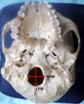
The study sample comprised 83 dry adult human crania. On the basis of the morphological characters as specified by Jain et al.(2013), 55 crania were found to be of males and 28 were of females (Satapathy and Sahoo, 2014). Only the skulls with no apparent deformity were included in the study (Figure 1). Initial examination of all the crania was done following the non-metric observations to categorize them into male and female categories. Data were collected from Department of Anthropology, Utkal University. Some direct and indirect measurements pertaining to foramen magnum were taken on each crania in accordance with the standard measurement techniques recommended by Martin and Saller (1957).
2.1 Definitions, Formula and procedures
Landmarks
Basion (ba): It is the point on the anterior border of the foramen magnum where it meets the median line.
Opisthion (o): It is the medial point on the posterior margin of the occipital foramen.
Length or antero-posterior diameter of foramen magnum (LFM) -It is measured as the straight distance between basion (ba) and opisthion (o).
Breadth or transverse diameter of foramen magnum (BFM) - It is measured as the maximum breadth of the occipital foramen transverse to the length line i.e. the median line.
Foramen magnum index= (Breadth of occipital foramen magnum/ Length of foramen magnum) *100
Area of foramen magnum is calculated from length and breadth of foramen magnum utilizing different formulae given by Routal(1984) and Tiexeria(1982) .
Formula given by Teixeria( 1982): Area = π ([LFM+BFM]/4)2
Formula given by Routal et al (1984): Area= LFM*BFM*π/4
Statistical analysis
The data were analysed using SPSS 20.0 program, the differences in mean were analyzed using t-test and the level of significance in the mean were analyzed at p<0.05 level.
3. Results And Discussion:
In the following tables the measurements on the skulls considered in the present study along with as to how these measurements compare with the corresponding ones obtained by different authors are presented. While Table 3.1 presents the mean values of the various measurements, the standard error mean and standard deviation with regard to both male and female skulls, Table 3.2 presents, respectively for females and males corresponding results of other workers from different regions.
Table 3.1: Descriptive statistics of the foramen magnum measurements for sample considered in the present study.
|
Measurements |
Male |
Female |
t-value |
||||
|
Mean |
S.E. |
S.D |
Mean |
S.E. |
S.D |
||
|
Length of foramen magnum (LMF) |
3.65 |
0.026 |
0.193 |
3.52 |
0.049 |
0.262 |
0.018 |
|
Breadth of foramen magnum(BFM) |
3.12 |
0.024 |
0.179 |
2.98 |
0.038 |
0.203 |
0.002 |
|
Foramen magnum index |
85.48 |
0.655 |
4.81 |
84.66 |
1.188 |
6.28 |
0.533 |
|
Area of foramen magnum (Teixeira) |
9.00 |
0.115 |
0.846 |
8.29 |
0.188 |
0.997 |
0.002 |
|
Area of foramen magnum(Routal) |
8.94 |
0.114 |
0.844 |
8.23 |
0.186 |
0.985 |
0.002 |
Table 3.2: Comparison of foramen magnum measurements with other studies
|
Sl. No |
Name of the worker |
Place and Country of Work |
Sample size of study |
Year of Work |
Length of Occipital Foramen (in cm) |
Breadth of Foramen (in cm) |
Foramen Index |
Area of The FM by Teixeria |
Area of the FM by Routal |
|||||
|
M* |
F |
M |
F |
M |
F |
M |
F |
M |
F |
|||||
|
1 |
Manoel, C. et al. |
Brazil |
139 males and 76 females |
2009 |
3.57 |
3.51 |
3.03 |
2.90 |
84.87 |
92.06 |
8.55 |
7.18 |
8.49 |
7.17 |
|
2 |
Ukoha U. et al. |
Nigeria |
90 males and 10 females |
2011 |
3.62 |
3.43 |
3.09 |
2.80 |
85.36 |
81.63 |
8.84 |
7.61 |
8.78 |
7.54 |
|
3 |
Singh G., Talwar I. |
Punjab University, Chandigarh, India |
26 males and 24 females |
2013 |
3.35 |
3.23 |
2.77 |
2.72 |
82.69 |
84.21 |
7.35 |
6.94 |
7.29 |
6.89 |
|
4 |
Santhosh, CS. Et al. |
Davangere, Karnataka |
63 males and 38 females |
2013 |
3.40 |
3.30 |
2.80 |
2.70 |
82.35 |
81.82 |
7.55 |
7.06 |
7.48 |
6.99 |
|
5 |
Tanuj Kanchan et al. |
Mangalore, Karnataka, India |
69 males and 49 females |
2013 |
3.45 |
3.36 |
2.73 |
2.67 |
79.13 |
79.46 |
7.50 |
7.31 |
7.40 |
7.04 |
|
6 |
Jain, S.K et al. |
Moradabad, U.P. |
70 males and 70 females |
2013 |
3.69 |
3.2 |
3.15 |
2.90 |
85.37 |
90.63 |
9.19 |
7.30 |
9.13 |
7.28 |
|
7 |
Jain Deepali et al. |
Punjab university & Delhi university, India |
70 males and 70 females |
2014 |
3.62 |
3.40 |
3.13 |
2.83 |
86.46 |
83.24 |
8.95 |
7.61 |
8.90 |
7.55 |
|
8 |
Present study |
Utkal university, vanivihar |
55 males and 28 females |
2014 |
3.65 |
3.52 |
3.12 |
2.98 |
85.48 |
84.66 |
9.00 |
8.29 |
8.94 |
8.23 |
*M- Male, F-Female
3.1 Discussion
The sex determination of incomplete or damaged skeletons is a difficult task in forensic science. In this effort, however, the foramen magnum plays a vital role because of its strength to remain intact while the rest of the cranium gets compromised (Graw, 2001). Further as the degree of sexual dimorphism within the foramen magnum reaches its adult size rather early in childhood (Scheuer and Black, 2004 ), it is therefore unlikely for it to respond to significant secondary sexual changes. As regard to its mechanical aspects, no muscle acts upon the shape and size of the foramen magnum, and its prime function is to accommodate the passage of structures into and out of the cranial base region, in particular to medulla oblongata, which occupies the greatest portion of the foramen space. In forensic investigation, sex determination is a necessary requirement for identification of skeletal remains in their advance state of decomposition.
It is interesting to observe the following from the information and the results displayed in Table-2,
-
The study on human skulls for sex determination is being carried out world over in different continents with different sample size.
-
LFM is invariably greater than BFM both for male and female skulls.
-
LFM (BFM) for male skulls is greater than LFM (BFM) for female skulls. But the difference is, in some samples, significant and in some samples not significant. For example, in Nigerian sample (Ukoha et. al., 2011), in the samples considered by Jain et al. (2013), Santhosh CS et al. (2013), and Deepali Jain et al. (2014) the differences both in LFM and BFM are significant, whereas in the samples considered by Gagandeep & Talwara (2013) and Kanchan et al. (2013) the differences both in LFM and BFM are not significant. Further in some samples difference in LFM and BFM need not be simultaneously significant or not significant. For example,in Brazilian sample (Manoel. et al.,2009) the difference in LFM is not significant but in BFM, it is significant. However, in the sample on which the present study is based the differences both in LFM and BFM are quite significant.
-
Cutting across the samples presented, it is observed that there are female skulls with LFM (BFM) greater than LFM (BFM) of male skulls. For example the mean LFM (BFM) of female skulls in the present study is greater than the mean LFM (BFM) of male skulls in the samples considered by Singh and Talwar (2013) or by Santhosh , CS et al. (2013) or by Kanchan et al. (2013).
-
Cutting across the samples presented, it has been observed that the area of foramen magnum of males shows a higher value as compared to females. However, the foramen index is not consistent between the sexes. For example,
4. Conclusion
There was morphometric difference but no morphological difference of the foramen magnum between genders. The study observes that the craniometric analysis of foramen magnum should be used only as supporting study in estimation of sex of fragmentary remains of skull, when other skeletal remains are preserved. The present study successfully identified the sex via foramen magnum in length, breadth and area. Index of the foramen magnum does not appear to be a very good variable for sex determination. Area of the foramen magnum is observed to have better accuracy in estimation of the sex of the skulls compared to the length and breadth of the foramen magnum. The present study is based on a limited sample. A study based on larger samples of documented Indian skulls needs to be undertaken for ascertaining the reliability of morphometric measurements of foramen magnum in sex determination.

Fig.1 Foramen magnum measurements: (LFM) maximum Length, (WFM) maximum width
References
- Babu Raghavandera, Y. P., Manjunathan, S., Rastogi, P., Kumar Mohan, T.S., Bhat, Virendra., Janardhan,Y.K., Kumar Pradeep, G., Kishan, K., and Dixit, P.N. (2011). Determination of Sex by Foramen Magnum Morphometry in South Indian Population. Indian J Forensic Med. Pathol., 41, 29-33.
- Deshmukh, A.G., and Devershi, D.B.(2006). Comparison of Cranial Sex Determination by Univarite and Multivariate Analysis. Journal Anat. Soc. India, 55(2), 48-51.
- Gopalkrishana, K., and Rathna, B.S.(2015). The craniometric study of foramen magnum of Indian population amd variations in its dimensions. Int. J. of Allied Med. Sci. and Clin. Research (IJAMSCR), 3(2), 205-211.
- Graw M. (2001). Morphometrische and Morphognostische. Geschlecthsdiagnostikan der menschlichenSchadelbasis.In Oehmicen M, Geserick G (eds) Osteologischeldentifikation and Altersschatzung Schmidt- Romild, Lubeck, pp. 103-121.
- Gunay, Y., Altinkok, M. (2000). The value of the size of foramen magnum in sex determination. J clin. Forensic med., 7(3), 147-149.
- Jain Deepali., Jasuja O.P., Nath, S.( 2014). Evaluation of foramen magnum in sex determination from human crania by using discriminant function analysis. Elective Medicine Journal, 2(2), 89-92
- Jain, S.K., Choudhary, A.K., Mishra, P.( 2013). Morphometric Evaluation of Foramen magnum for sex determination in a documented North Indian sample. Journal of Evolution of Medical and Dental Sciences, 2(42), 8093-8098.
- Kanchan, Tanuj., Gupta, Anadi., and Krishan Kewal.(2013), Craniometric Analysis of Foramen Magnum for Estimation of sex. International journal of Medical, Health, Biomedical, Bioengineering and Pharmaceutical Engineering, 7(7), 111-113.
- Krogman, W.M., Iscan, M.Y. (1986). The Human skeleton in forensic medicine Springfield, Illinois: Charles Thomas pub. Ltd.
- Manoel,C., Prado, FB., , Caria, PHF., and Groppo, FC. (2009). Morphometric analysis of the foramen magnum in human skulls of Brazilian individuals: its relation to gender. Braz. J. Morphol. Sci., 26(2), 104-108.
- Macaluso Jr., PJ.(2011). Matric sex determination from basal region of the occipital bone in a documented French sample. Bull. Mem Soc Anthropol. Paris, 23,19-26.
- Martin, R., and Saller, K. (1957). Lehrbuch der anthropologie Stuttgart: G. Fischer.
- Radhakrishna, S.K., Shivarama, C.H., Ramakrishna, A., and Bhagya, B. (2012). Morphometric analysis of Foramen magnum for Sex Determination in South Indian population. Nitte University. Journal of Health Science, 2(1), 20-22.
- Routal, R. V., Pal, G.P., and Bhagwat, S.S. (1984). Relationship between endocranial volume and the area of the foramen magnum. J. Anat. Soc. India, 33 (3), 145-149.
- Santosh CS., Vishwanathan, KG., Gupta, A., Siddesh, RC., and Tejas, J.(2013) Morphometry of the Foramen Magnum; An Important Tool in Sex Determination. Research and Review Journal of Medical and Health Sciences, 2(4), 88-91.
- Satapathy KC and Sahoo Bidyabharati.( 2014). Estimation of Endocranial Capacity and Identification of Sex from Adult Human Skull of Eastern India.; The Tribal Tribune, 7(1). Available from www.etribaltribune.com.
- Scheuer, L and Black, S. (2004). The Juvenile Skeleton San Diego, CA: Elsevier Academic Press.
- Uysal,S., Gokharman, D., Kacar,M., Tuncbilek, I., and Kosa, U. (2005). Estimation of sex by 3D CT measurements of foramen magnum. J Forensic Sci., 50(6),1310-1314.
- Singh, Gagandeep., and Talwar, I.( 2013). Morphometric analysis of foramen magnum in human skull for sex determination. Human Biology Review, 2(1), 29-41.
- Suazo, GIC., Russo, PP., Zavando, MDA., and Smith, RL.(2009). Sexual dimorphism in foramen magnum dimensions. International Journal of Morphology, 27 (1), 21-23
- Ukoha U., Egwu, OA., Okafor, IJ., Anyabolu, AE., Ndukwe, GU., and Okpala, I.( 2011). Sexual Dimorphism in the Foramen Magnum of Nigerian Adult. International journal of Biological & Medical Research, 2(4), 878-881.
- Teixeira, WR.(1982).Sex identification utilizing the size of Foramen Magnum. American Journal of Forensic Medicine and Pathology, 3(3), 203-206.
- Wescott, DJ., and Moore- Jansen, PH.(2001). Metric variation in the human occipital bone: forensic anthropological applications. Journal of Forensic Sciences, 46(5), 1159-1163.
- Dr. K. C. Satapathy, Reader, Dept. of Anthropology, Utkal University, Bhubaneswar, Odisha
- Bidyabharati Sahoo, Research Scholar, Dept. of Anthropology, Utkal University, Bhubaneswar, Odisha


